Review Article
Year: 2023 |Volume: 2 | Issue: 01 |Pages: 01-07
Comparative Structural Study of Asthi, Peshi and Snayu for The Purpose of Bharsahatwa WSR to Sushrut Samhita.
About Author
Correspondence Address:
Dr. Manish Ashok Umak PG sch. Rachana Sharir RTAM Akola.
Date of Acceptance: 2022-12-17
Date of Publication:2022-01-11
Article-ID:AYU_75_01_23 https://ayuscript.com
Source of Support: Nill
Conflict of Interest: None declared
How To Cite This Article: Umak M.A., Deshpande Y.N., Kandekar S.M., Pimparkar K.M. Comparative Structural Study of Asthi, Peshi and Snayu for The Purpose of Bharsahatwa WSR to Sushrut Samhita.AYUSCRIPT 2023;2(1):1-7
Abstract
According to Ayurveda Asthi, Peshi, Snayu are the factors in human body which supports body posture and locomotion, but according to Acharya Sushrut only Snayu which are 900 in number are said to be responsible for Bharsahatwa i.e., Weight Transmission. Asthi is called Bone, Peshi is called Muscles & Snayu is called Tendon. Bones are suppose to be the strongest part of body and are responsible for posture and locomotion. Bones also help in protection of vital organs such as Brain, Heart. Muscles help in body movement by contracting and relaxing themselves which results in movement of body parts. In this present study we are going to prove that the statement given by Acharya Sushrut i.e. Varoius Ligament present in body are responsible for weight transmission, in Sushrut Samhita is true. For this study we have compared histology of Bones, Muscles & Tendons. Result shows that Histologically Tendons are more rigid, strong & responsible for weight transmission than Bone & Muscles with the help of comparison of histology of Asthi, Peshi & Snayu.
KEYWORDS: – Asthi; Peshi; Snayu; Bone; Muscles; Tendon; Bharsahatwa; Weight Transmission
Introduction
Histology of Bones1:
Bone is a tissue in which the extracellular matrix is hardened to work as a supporting function. There are three key cells of bone tissue which are Osteoblasts, Osteocytes and Osteoclasts. They each have unique functions and are derived from two different cell lines.
- Osteoblasts synthesize the bone matrix and are responsible for its mineralization. They are derived from osteoprogenitor cells, a mesenchymal stem cell line.
- Osteocytes are inactive osteoblasts that have become trapped within the bone they have formed.
- Osteoclasts break down bone matrix through phagocytosis. They are supposed to be derived from the monocyte (macrophage) cell line.
Aim: -
To prove Ligament present in body are responsible for weight transmission.
Objectives-
1 To study structural details of Asthi i.e., Bones.
2. To study structural details of Peshi i.e., Muscles.
3. To study structural details of Snayu i.e., Ligaments.
4. To compare the above structures in the view of weight transmission property.
Research question – Which structure is responsible for weight transmission of body, whether it is Bone or Muscle or Ligament?
The balance between osteoblast and osteoclast activity maintains bone density and ensures that bone is neither overproduced nor over degraded. These cells build up and break down bone matrix.
Bone is divided into two types which are different structurally and functionally.
- Compact bone or cortical bone: - These mainly serve for mechanical function. In this area of bone ligaments and tendons attach. It is thick and dense in structure.
- Trabecular bone or cancellous bone or spongy bone: - These bones mainly serves a metabolic function. This type of bone is located between layers of compact bone and is thin and porous in structure. Which is located within the trabeculae of the bone marrow.
- Muscles are multicellular contractile units. They are divided into three types: Skeletal muscle, Smooth muscle, Cardiac muscle.
- Skeletal Muscle: - Skeletal muscle is mainly responsible for the movement of the skeleton, it is also found in organs such as the eye ball and the tongue. These are voluntary muscles, and so are under conscious control.
-
The components and structures of skeletal muscle cells are similar as other cells but different terms are used to describe those muscle cells. The plasma membrane of skeletal muscle is called the sarcolemma, its cytoplasm is known as sarcoplasm, and the endoplasmic reticulum is called the sarcoplasmic reticulum.
Each muscle cell is defined by a sarcolemma and contains many nuclei displaced peripherally along its length. While at the centre a large number of longitudinal myofibrils, groups of arranged contractile proteins are present.
The myofibril contains many important histological landmarks: The myofibril is composed of alternating bands. The I-bands (isotropic in polarized light) are light in color and the A-bands (anisotropic in polarized light) are dark in color. The alternating pattern of these bands results in the striated appearance of skeletal muscle.
Each myofibril can be defined as a series of contractile units called sarcomere that contains two types of filaments: thick filaments which are composed of myosin, and thin filaments which are composed of actin.
These separate filaments do not change in length during muscle contraction, but the thin filaments slide over the thick filaments to shorten the sarcomere.
Skeletal muscles are divided into two muscle fiber types:
-
- Slow-twitch (type I) muscle fibers contract slowly and it rely on aerobic metabolism.
- Fast-twitch (type II) muscle fibers contract rapidly due to the presence of a faster myosin.
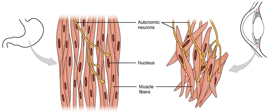
- Smooth Muscle: - Smooth muscle mainly forms the contractile portion of the wall of the digestive tract from the middle portion of the esophagus to the internal sphincter of the anus. Smooth muscles are controlled by autonomic nervous system, hormones and local metabolites. Since it is not under conscious control, smooth muscle is involuntary muscle. Smooth muscle fibers are elongated spindle-shaped cells with a single nucleus. The contractile proteins of these cells are not arranged into myofibrils like those of skeletal and cardiac muscle, so they appear smooth and not striated.
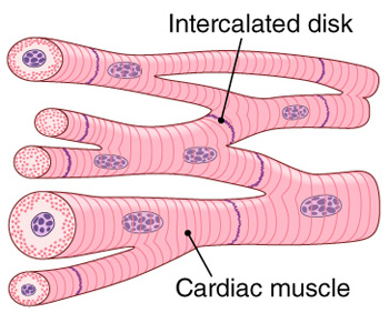
- Cardiac Muscle: - Cardiac muscle shares important characteristics like both the skeletal and smooth muscle. Cardiac muscle produces strong contractions like skeletal muscle, but it shows property to initiate continuous contraction like smooth muscle. The rate and force of contraction is same as muscle with voluntary control, but is influenced by the autonomic nervous system and hormones. Histologically, cardiac muscle appears striated like the skeletal muscle due to arrangement of contractile proteins.
Histology of Ligament8:
Ligaments and tendons are soft collagenous tissues. Ligaments connect bone to bone and tendons connect muscles to bone. Ligaments and tendons play a significant role in musculoskeletal biomechanics. They have an important role in orthopedic treatment. As like all biological tissues, ligaments and tendons have a hierarchical structure that affects their mechanical behavior. Also ligaments and tendons can adapt due to changes in their mechanical environment because of injury, disease or exercise. Thus ligaments and tendons are another example of the structure-function concept and the mechanically mediated adaptation concept that permeate this biomechanics course.
Ligaments and tendons have a hierarchical structure.
The largest structure in the above image is the tendon (shown) or the ligament. The ligament or tendon then is split into smaller entities called fascicles. The fascicle contains the basic fibril of the ligament or tendon, and the fibroblasts, which are the biological cells that produce the ligament or tendon. The structural characteristic at this point that plays a significant role in the mechanics of ligaments and tendons is the crimp of the fibril. The crimp is structure with waviness of the fibril; this contributes significantly to the nonlinear stress strain relationship for ligaments and tendons and for basically all soft collagenous tissues.
Components:
- Components of Bone10: two main components of bone are collagen and calcium phosphate, These distinguish it from other hard tissues such as chitin, enamel, and shell.
- Components of Muscle11: An individual skeletal muscle is composed of hundreds, or even thousands of muscle fibers bundled together and which are wrapped in a connective tissue covering. Each muscle is surrounded by a connective tissue sheath which is called the epimysium. Fascia a connective tissue outside the epimysium, surrounds and separates the muscles.
- Components of Ligament12: and ligaments are composed of type I collagen fibers which are surrounded by a mesh of loose connective tissue. The whole tendon transmits forces from muscle to bone. It also shows viscoelastic behavior such as creep or stress relaxation.
Discussion
Comparison of structural study of Bone, Muscles & Tendon
In above project we have studied histology and macroscopic details of above structures. The study suggest that
-
-
- Bone has very strong cellular structure, unit of it is osteocyte. Physiologically the cells are mainly made up of calcium & other minerals so they are very tough in nature. They are mainly useful for protection of vital organs & locomotion, but bones have not any specialized structure which joins it to other bone, so it is not responsible for weight transmission.
- Muscles are made of specialized protein tissue, which attach the 2 bones. By contraction & relaxation movement they are responsible for movement of joints. So due to muscular action various movements of locomotion take place. But the structure of muscle is soft and weak so they can not attach 2 bones in joints. So, it can be said that muscles are responsible for carrying of weight by motion of joints but not responsible for weight transmission.
- In case of Ligament, the structural study shows that, the tissue of ligament is made in such a way that they are tougher than muscular tissue, but slight soft than bony tissue. Moreover, the ligaments can perform flexion & extension movement so that they can easily move the joint.
-
Specialty of Ligaments is their location. They are located in the synovial cavity of joint; at this location they firmly hold the articulating surfaces of bones at their places. Furthermore, if the ligament is damaged by any trauma, they can redevelop themselves for their function. So due to all these properties Ligaments which are also called Snayu are responsible for weight transmission.
Conclusion
The comparison between Bone, Muscle & Tendon shows that Histologically Tendons are more rigid strong & responsible for weight transmission than Bone and Muscle. So we could conclude that the statement given by Acharya Sushrut is accurate.
References
- No title [Internet]. Medcell.org. [cited 2022 Nov 20]. Available from: http://medcell.org/histology/bone_lab.php
- Structure of bone tissue [Internet]. Cancer.gov. [cited 2022 Nov 21]. Available from: https://training.seer.cancer.gov/anatomy/skeletal/tissue.html
- No title [Internet]. Medcell.org. [cited 2022 Nov 20]. Available from: http://medcell.org/histology/muscle_lab.php
- Histology of muscle [Internet]. Etsu.edu. [cited 2022 Nov 21]. Available from: https://faculty.etsu.edu/forsman/histologyofmuscleforweb.htm
- Lecturio.com. [cited 2022 Nov 21]. Available from: https://cdn.lecturio.com/assets/Smooth-muscle-tissue-types.jpg
- Onlinebiologynotes.com. [cited 2022 Nov 21]. Available from: https://www.onlinebiologynotes.com/wp-content/uploads/2018/02/cardiac-muscle.jpg
- Jaishree G. Differentiate between Striated, Smooth and Cardiac Muscles on the basis of their structure and Site/ Location in the body [Internet]. CBSE Class Notes Online - Classnotes123. 2021 [cited 2022 Nov 21]. Available from: https://classnotes123.com/differentiate-between-striated-smooth-and-cardiac-muscles-on-the-basis-of-their-structure-and-site-location-in-the-body/
- Structure and Function of Ligaments and Tendons [Internet]. Wsu.edu. [cited 2022 Nov 21] Available from:- http://sites.bsyse.wsu.edu/pitts/be120/Handouts/animal%20tssue%20descriptions%20and%20mechanical%20proprties.htm
- Researchgate.net. [cited 2022 Nov 21]. Available from: https://www.researchgate.net/figure/FIGURE1-The-structural-hierarchy-of-ligament-and-tendon-Adapted-from-Kastelic_fig1_11626143
- Heaney RP, Donald Whedon G. bone. In: Encyclopedia Britannica. 2022.
- Structure of skeletal muscle [Internet]. Cancer.gov. [cited 2022 Nov 21]. Available from: https://training.seer.cancer.gov/anatomy/muscular/structure.html
- Zschäbitz A. Anatomie und Verhalten von Sehnen und Bändern. Orthopade [Internet]. 2005 [cited 2022 Nov 21];34(6):516–25. Available from: https://pubmed.ncbi.nlm.nih.gov/15926082/